Dermoid Cyst, also known as “Mature Cystic Teratoma”, is a type of benign ovarian germ cell tumor from the gynecologists’ point of view. The expression of “from the gynecologists’ point of view” must be emphasized because dermoid cysts may emerge from many parts of the body including the skin, brain and even the eyes (1).
However, ovarian dermoid cysts differ from dermoid cyst situated in other organs, in terms of structure and development. Orbital (relating to the eyeball) dermoid cysts develops in consequence of cyst formation by “aberrant ectodermal” tissues, whereas ovarian dermoid cysts differ form them and originate from each of the three germ layers that play a role in the formation of the embryo. (1, 2)
If the last sentence needs to be more clarified, ovarian dermoid cysts originate from all three layers (endoderm/mesoderm/ectoderm), which play a role during embryonic development.
Dermoid cysts of the ovary constitute 10-20% of all ovarian tumors (all including any benign and malignant ones). It is the MOST COMMON ovarian germ cell tumor and the ovarian tumor MOST COMMONLY seen in people under 20 years of age (3, 4).
Ovarian Germ Cell Tumors; what is the Group that Dermoid Cysts are Classified in?
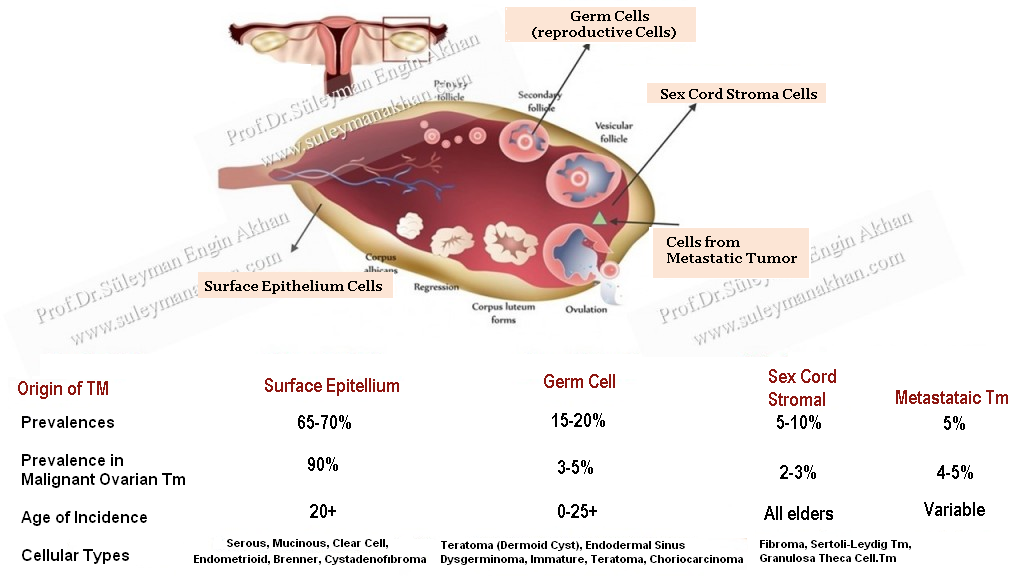
In the following figure, you see which tumor origins from which tissue of the ovary. The classification describes tissues that all benign and malignant tumors originate from, by means of a simple diagram. As is seen, mature cystic teratomas originate from the reproductive cells, and consist of mature cells as the name implies.
Now, let me share with you an important secret. 🙂 No matter whether in humans or any other living creatures, the more the tumoral tissues have resemblance to normal cells, the more they act as benign tumors; i.e, they do not metastasize in an incoherent way and do not recur, no matter the extent of the maturity, i.e. abnormality of the cells of the consisted tomoral tissue. After all, they are benign.
Therefore, the word “mature” indicates that important and benign dermoid cysts are often benign. If we call them “immature”, they are malignant. For example, immature teratomas malignant tumors.
As you can also see on the chart, dermoid cysts (mature cystic teratomas) constitute a sub-group of teratomas among germ cell tumors (reproductive cell tumor tumors).

If we need to go into detail, ovarian germ cell tumors are classified in two main groups:
a. Embryonal tissue-derived tumors (teratomas, their subgroups, and disgerminomas)
b. Tumors originating from extra-embryonic fetal cell populations (e.g. placenta), and tumors formed jointly by cells of the both groups.
A significant part of these neoplastic formations, especially the ones that constitute the teratoma part, have a benign structure. They are not malignant. If they need to be classified according to the World Health Organisation (5):
- Teratomas: Teratoma means “Monster Tumor” in Greek To tell the truth, although teratomas are usually benign, they deserve this characterization in the sense of their appearance and content.
a. Benign cystic mature teratomas; Dermoid Cysts. The MOST COMMON ovarian germ cell tumors are dermoid cysts.
b. Immature teratoma: They are malignant germ cell tumors. - Dysgerminoma: It is the female version of seminoma (testicular tumor) seen in males. It consists of immature germ cells. The MOST COMMON malignant germ cell tumor seen in women is ovarian tumor. It is also the MOST COMMON malignant ovarian tumor in pregnants (6). Between ourselves, the patient can get pregnant with a good surgical treatment, and there is no need to remove the uterus and the ovary that does not contain any tumor. LDH (Lactate Dehydrogenase Enzyme) can be used as a tumor marker.
- Endodermal sinus (yolk sac) tumor: They are cancers caused by the structure that comes into existence next to the embryo during the first weeks of pregnancy, secretes progesterone, and bears germ cells. They are rarely seen. Alpha-FetoProtein (AFP) is used as a tumor marker. Patients with high AFP levels are more likely to be diagnosed with endodermal sinus tumor.
- Mixed Germ Cell Tumors: They are typically composed of the combinations of Teratoma+Yolk sac tumor (endodermal sinus tm), dysgerminoma+teratoma, and/or embryonal carcinoma.
- Very Rare Ovarian Germ Cell Tumors: Pure embiyonel cancers, non-gestational Choriocarcinoma, Pure polyembryoma
As is seen, dermoid cysts are classified under the title of “Teratoma”; and they are benign tumors. Dermoid cyst diagnosed after 40-45 years of age can be malignant.
Malignant ovarian germ cell tumors have always attracted my interest because they are commonly seen in young women.
Therefore, I have alsa carried out academic studies. 🙂 If you want to read my article published in the international journal SCI,
please click on the following link: http://www.ncbi.nlm.nih.gov/pubmed/21319513
References
1. http://emedicine.medscape.com/article/1218740-overview
2. http://emedicine.medscape.com/article/1112963-overview
3. Stany MP, Hamilton CA. Benign disorders of the ovary. Obstet Gynecol Clin North Am. 2008; 35(2):271-284.
4. Wu RT, Torng PL, Chang DY, et al. Mature cystic teratoma of the ovary: a clinicopathologic study of 283 cases. Zhonghua Yi Xue Za Zhi (Taipei). 1996; 58(4):269-274.
5. Tavassoli FA, Devilee P, eds. World Health Organization Classification of Tumors: Pathology and genetics of tumours of the breast and female genital organs. Lyon, France: IARC Press; 2003
6. Gordon A, Lipton D, Woodruff JD. Dysgerminoma: a review of 158 cases from the Emil Novak Ovarian Tumor Registry. Obstet Gynecol. 1981;58(4):497–504
